3d live cell imaging
Patented Cell-CT Imaging High-resolution 3D images Cell-CT of individual cells contained in the sputum measure hundreds of disease indicators and molecular markers in each cell. 88 million by 2027.

Live Fucci Mesc Imaged For Over 48 Hours With Nanolive S 3d Cell Explorer Fluo 3d Cell Cell Free Labels
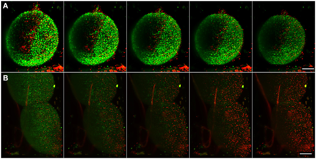
3D subcellular intravital imaging in mammals with low phototoxicity.

. The EVOS M5000 microscope is a workhorse for cell imaging labs across a broad range of applications and imaging requirements. State-of-the-art subcellular IVM in mammals such as resonant scanning two-photon microscopy and SDCM captures only in-focus 2D information with unnecessary. The hierarchical model of chromatin organization has been.
DNA accessibility depends on chromatin structure and dynamics which basically control all DNA-related processes such as transcription DNA replication and repair. High-speed 3D imaging with multi-site DAO thus provides a robust and accurate tracking analysis for various organelle dynamics. Equipped with a brightfield and two fluorescence channels green and red the CytoSMART Omni FL can be used for continuous live-cell imaging as well as endpoint assays.
Organoid cultures from tissue to mature 3D organoid culture and their downstream analysis. Emotions coordinate our behavior and physiological states during survival-salient events and pleasurable interactions. 18 million in 2021 and expected to reach USD 2775.
THUNDER Imagers with Computational Clearing define a new class of instruments for high. From endpoint and live-cell imaging applications to imaging and analyzing 2D monolayers 3D cell models and organ-on-a-chip platforms the world of cellular imaging is very diverse Download eBook. Many proteins that express to the surface of cells are targets for the discovery and development of biopharmaceuticals.
The CubiX product line is the sum of the next-generation 3D cells culture technologies. Transform your cell imaging. To answer important scientific questions they enable you to obtain a clear view of details even deep within an intact sample in real time without out-of-focus blur.
Based on our patented. With a high-resolution CMOS monochrome camera capable of four-color. Sharp imaging of 3D specimens is now as easy as working with your favorite camera-based fluorescence microscope.
For instance G-protein coupled receptors GPCRs are the largest class of cell-surface proteins and are targets for almost 40 of existing drugs. 37 during the forecast period with market size of USD 1802. Even though we are often consciously aware of our current emotional state such as anger or happiness the mechanisms giving Emotions are often felt in the body and somatosensory feedback has been proposed to trigger conscious emotional experiences.
The inverted microscope provides 125x to 60x magnification in fluorescence brightfield and color brightfield while the upright microscope enables other common applications including ELISpot slide. Cytation C10 provides the perfect environment to grow and analyze live cells over time. A Petri dish a 3D cell culture allows cells in vitro to grow in all directions similar to how they would in vivo.
This webinar will demonstrate the use of hepatic organoid cultures to validate a label-free live-cell toxicology screen using a powerful and flexible Incucyte Live-Cell Analysis System. A 3D cell culture is an artificially created environment in which biological cells are permitted to grow or interact with their surroundings in all three dimensions. It enables long-term culture of human tissues biopsies 3D cell models or organ-on-a-chip models.
How the 2-m-long genomic DNA is packaged into chromatin in the 10-µm eukaryotic nucleus is a fundamental question in cell biology. The CERO 3D Incubator Bioreactor is a new revolutionary instrument creating optimal cell culture environment by monitoring and controlling temperature pH and CO2 levels. Our AI-based classification of cells can identify 1000 cell images per second and over 800 3D cell features at a 200 nanometer isotropic resolution.
Powerful movie maker and kinetic analysis software tools allow visualizing and analysis time-lapse experiments. These three-dimensional cultures are usually grown in bioreactors small capsules in. DISCOVER MORE ABOUT US.
Successful live cell kinetic imaging relies on a consistent environment including temperature control and CO 2 O 2 control and monitoring. Combining 3D Cell Culture Assays with Live Imaging Watch Now. The CytoSMART Omni FL is a live-cell imager capable of producing high-quality whole-well brightfield or high-throughput fluorescence images of living cells.
Semi-automation powerful software tools and a user interface designed by biologists for biologists means that the M5000 can revolutionize your imaging workflow. Real-time kinetic analysis of organoids. It enables ultra-fast temperature shifts in the 5-45C range while live-imaging cells.
Unlike 2D environments eg. 1- 4 individually controlled CEROtubes with a volume of up to 50ml provide highest biomass yields in a standardized and reproducible way with minimum handling requirements. Combining 3D Cell Culture Assays with Live Imaging Hear from Brad Larson Principal Scientist at BioTek Instruments Inc about how to combine the Cytation 5 with Corning spheroid high content microplates to easily perform assays simplify 3D spheroid workflow.
Discovery and selection of high value clones with elevated cell surface expression of GPCRs from a transfected pool of. Cytation 7 Cell Imaging Multi-Mode Reader combines automated digital upright and inverted widefield microscopy with monochromator-based multi-mode microplate reading. The Live Cell Imaging market is projected to register a CAGR of 7.

Trakine Pro Live Cell Tubulin Staining Kit Your Best Choice For Cytoskeleton Study Abbkine Antibodies Proteins Biochemicals Assay Kits For Life Science Research

Nanolive Imaging Nanolive A Complete Solution For Your Label Free Live Cell Imaging Free Labels Nuclear Membrane 3d Cell

3d Live Cell Time Lapse Compilation Cell Video Cell 3d Cell

The Most Powerful Solution To Explore Living Cells 3d Cell Cell Solutions

Label Free Live Cell Imaging In 3d Mitosis Of Mesc 3d Cell Cell Mitosis

Application Note Growing And Filming Stem Cells With The 3d Cell Explorer 3d Cell Stem Cells Application Note

Smartphones Used For Live Cell Imaging Imprimante 3d Impression 3d Microscopes

Pin Auf 3d Microscope Cell Images

Holographic 3d Cell Explorer Steve Set To Disrupt Cell Imaging Video 3d Cell Cell Holographic

Stem Cells Long Term Live Imaging Of Mouse Embryonic Stem Cells For 48 Stem Cells Cell Video 3d Cell

Stem Cells Long Term Live Imaging Of Mouse Embryonic Stem Cells For 15 Hours 3d View

Nanolive The Future Of Living Cell Microscopy

A Research Group Led By Kobe University S Professor Matoba Osamu Organization For Advanced And Integrated R Holography Cell Processes Nobel Prize In Chemistry

Multispectral Quantitative Phase Imaging Captures Live Human Cells Quickly Capture Systems Biology Cell


